Services
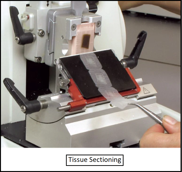
Microtomy (Tissue Sectioning)
Precision Tissue Sectioning for Detailed Analysis
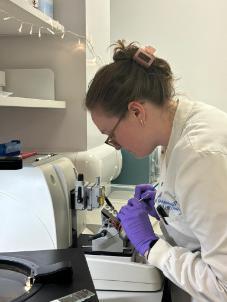
Using the Leica RM2255, GTRF provides ultra-thin sectioning of Formalin-Fixed Paraffin-Embedded tissues (FFPE) for downstream applications like histology and Immunohistochemistry (IHC) staining.
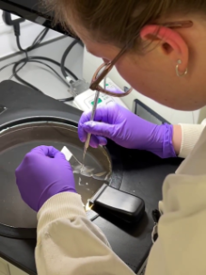
Key Features:
- Consistent section thickness for reproducible results.
- Expertise in handling diverse tissue types.

Brightfield & Fluorescence Scanning
Advanced Whole-Slide Scanning for Tissue Research
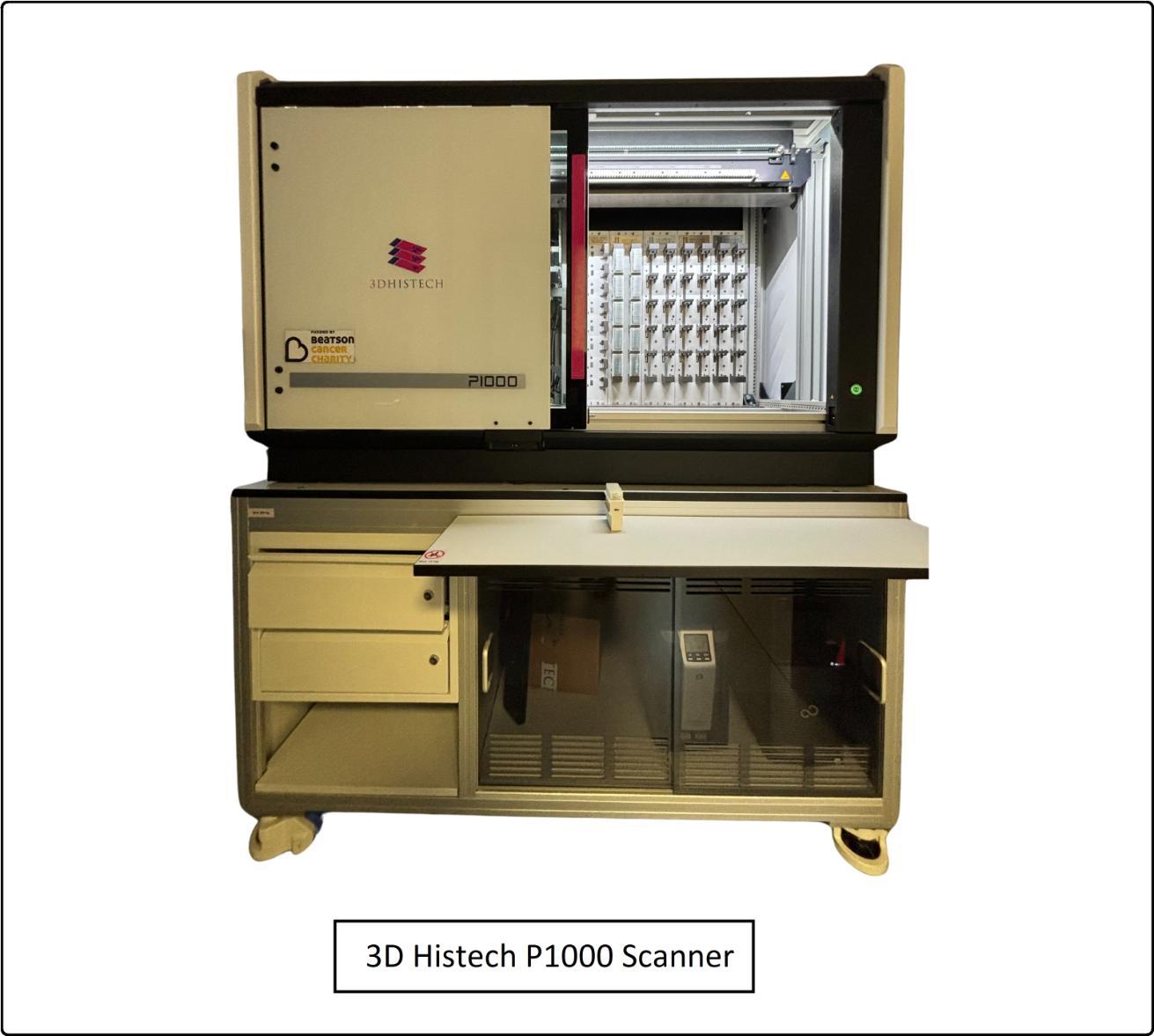
1. 3D Histech P1000:
- Capable of Brightfield scanning 1000 slides, including 100 megaslides and 900 standard slides.
- Scans at 20x/40x magnification in as little as 23 seconds per slide.
- Funded by Beatson Cancer Charity, this cutting-edge scanner enables rapid digitization of brightfield microscopy slides.
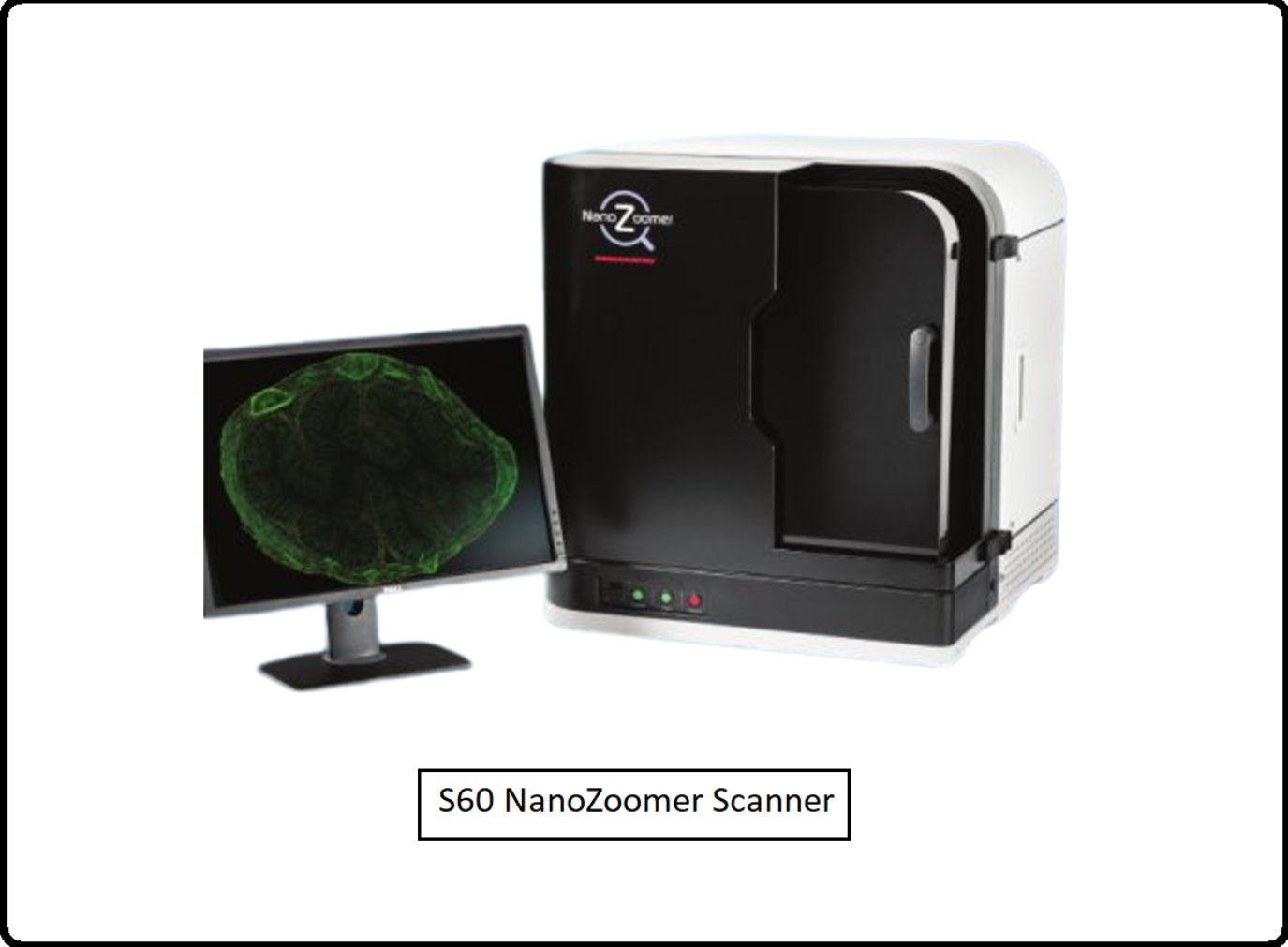
2. S60 Hamamatsu Nanozoomer:
- Capable of scanning 60 slides, including 10 megaslides and 60 standard slides.
- Scans up to 6 fluorescent channels including DAPI, at 20x/ 40x magnification capturing intricate multi-color details.
- Ideal for Fluorescence Scanning.
Data Management:
- Images securely stored on University of Glasgow’s dedicated servers.
- Accessible using the free NDP.view2 from Hamamatsu.
- Data sharing supported through Data Transfer Agreements (DTA).
Tissue Microarray Construction (TMA)
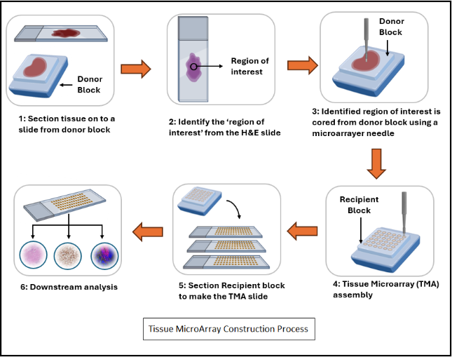
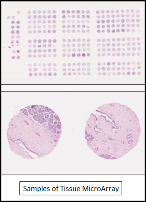
Customized TMA Solutions for High-Throughput Analysis
- Bespoke TMAs created for individual or shared research needs.
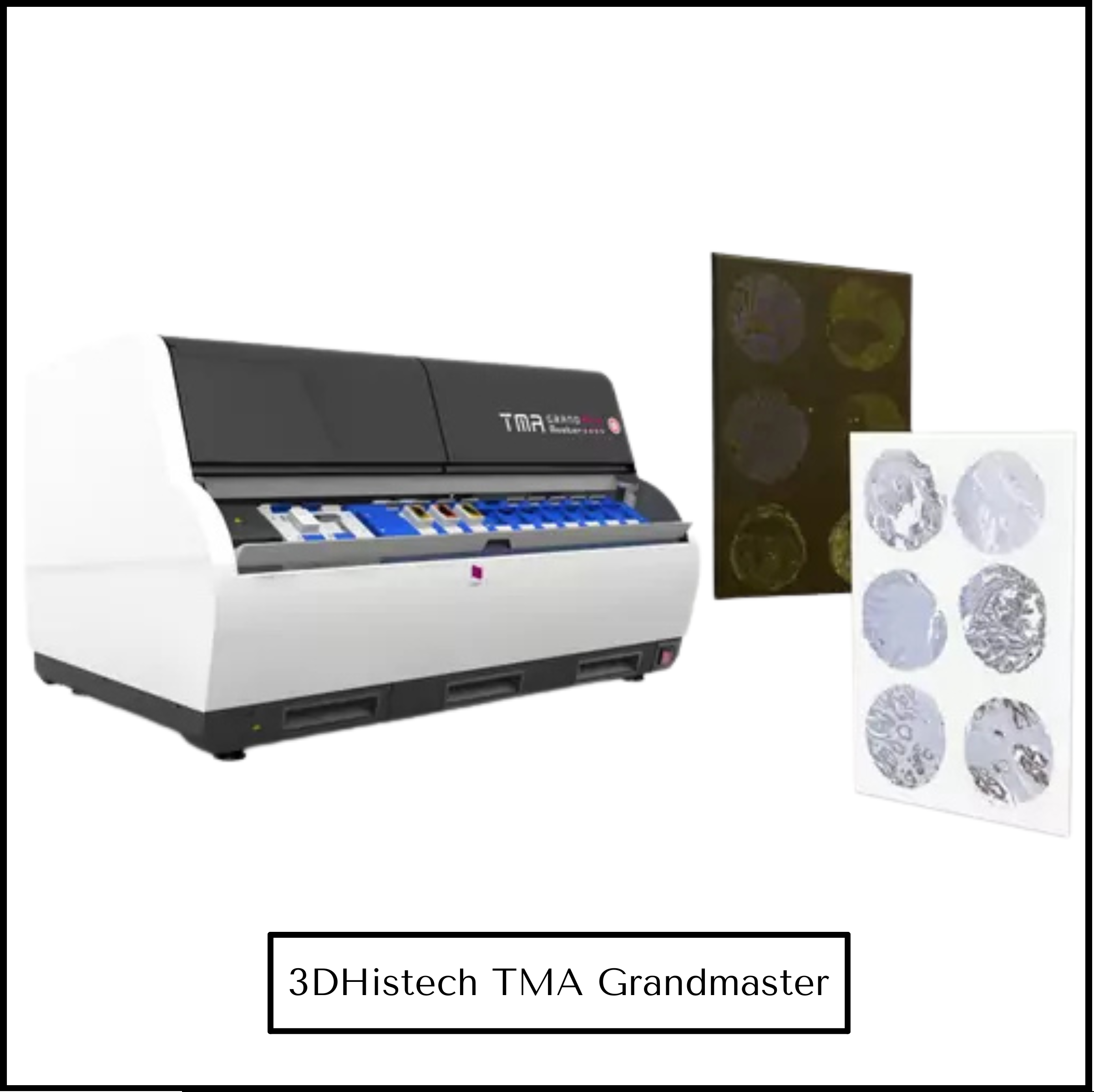
- 3DHISTECH TMA Grandmaster – By using TMA Grand Master you can easily and quickly create TMA blocks with great precision, of 0.6, 1, 1.5 and 2 mm diameter tissue cores. Then, the extracted tissue samples can be used later for various applications at the field of molecular pathology. TMA Grand Master prepares TMA blocks from biological specimens and samples of human or animal tissues.
- Constructed from ethically approved cases via the NHS Biorepository, allowing many samples in one TMA.
Advantages:
- Efficient analysis of multiple tissue samples on a single slide.
- Flexible custodianship of blocks and sections.
